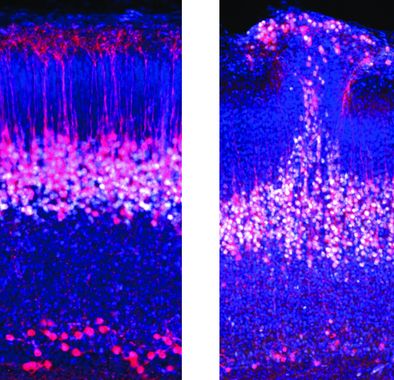
Image: Layer 5 pyramidal neurons in normal mice (left) compared with mice with autism gene knocked-out (right), showing a patch of disorganized cortex.
Congratulations to both of you! Could you tell us a bit more about what your newly published study is about?
Arjun Bharioke: Thanks! This study provides the first characterization of a specific class of cortical neurons, all the way from the first time when these neurons enter the developing cortex. We developed a method to image from these neurons during embryonic development, in living embryos, which therefore means that, for the first time, we can identify that neurons form embryonic circuits right when they first enter the cortex. This was quite different from what we had expected, when we first started this project, where we had thought that these neurons would only slowly develop activity, and then slowly form circuits.
Martin Munz: Fundamentally, the idea is to understand how neuronal circuits develop in as much detail as possible. We wanted to understand: When do neurons form the first synapses? When do they have action potentials? What is the cell type composition as the circuit forms? At which point do the neurons migrate? When do the neurons of a circuit start to communicate? Some of these questions can arguably only be addressed in vivo and all of this happens during embryonic development. By using our method of watching circuit development in the living mouse embryo, we were able to get an understanding of each of these processes in the normal circuit. Once we felt we had a good understanding of what is going on, we also wanted to see how neuronal circuits develop in a disease situation. Thus, we also looked at circuit development in mouse models of autism – a common neurodevelopmental disorder.
What are cortical circuits and why is it important to study them?
M: The cerebral cortex mostly consists of the six-layered neocortex, with its axonal connections that send information within cortex and to other brain areas. It plays a key role in awareness, attention, perception, thought, memory, language, and consciousness. The computation of all of this happens in neuronal circuits – ensembles of neurons that are the building blocks of each computation, or action. Therefore, it is important to understand how it works and how it develops.
A: The things that we believe define who we are – our intelligence, our morality, conscious perception – are formed, somehow, within the cerebral cortex. Cortical neurons form into circuits and – depending on their inputs, circuit organization, and outputs – these circuits somehow perform complex computations. However, amazingly, although we understand the biophysics and molecular biology of individual cortical neurons in great detail, the way in which circuits perform these computational transformations remains a mystery. There is no single cortical circuit where we could have an intuitive understanding of how it generates its functional role. Hence, understanding how cortical circuits function remains a crucial question in neuroscience.
How did you conduct your research and how long did it take to finish the project?
M: I started the project in 2015 when I started in the Roska lab. In 2016, Arjun joined the project because of a common interest in neuronal communication. We spent the next 3 years developing a method to image neurons in the living embryo using microscopy.
A. Indeed, it was pretty much three years of trial and error to finally figure out how to keep the embryos healthy and stable. That meant that, for almost three years, we didn’t have a single recording that was actually usable. Once we got the method working, then we managed to collect most of the data within the paper within the next two years. And we’ve spent just over a year and a half working on publishing the paper, including reviews, additional experiments etc. So, together, it’s been about 6 and a half years or so?
M. Yeah, 6 and a half years. However, we’ve been fortunate to be able to collaborate with other members of the Roska lab and others to answer different aspects of our questions. Hence, the project itself and the collaborations have made it a very interesting and fun 6 years.
What were your main findings?
A: I think the findings break into three parts. The first is the technical achievement of actually being able to make consistent recordings from cortical neurons, in living embryos. This is something that we hope will go into widespread use within the developmental neuroscience community. Second, we found that one specific type of cortical neuron, layer 5 pyramidal neurons, forms a new form of temporary circuit, right at the beginning of the formation of the cortex. This was unexpected, and it will be interesting to see if this is true for other cortical cell types, or if this is specific to this particular type of neuron.
M: Third, our findings relate to the question of how mutations in genes associated with autism- spectrum disorder in humans may affect the formation of embryonic cortical circuits. We found a disruption of the development of layer 5 pyramidal neurons when we mutate two different autism-associated genes. This suggests, to understand the way in which disorders like autism form, we can’t ignore the early circuits forming in embryonic development.
A. And that again brings it back to novel method of embryonic cortical recordings, which should prove valuable in the future study of disorders like autism spectrum disorder, in the future.
What is the significance of your findings for autism research?
M: Most generally, understanding how neuronal circuits form, step-by-step, allows us to determine at which point development differs in an animal model of autism. This might give us a better understanding of how autism develops in humans as well.
A: Additionally, our results suggest that changes to the layer 5 pyramidal neuron circuitry may be one of the components underlying at least part of the circuit changes associated with autism spectrum disorder. Which makes it crucial to study both these and other embryonic circuits.
What were your personal favorite moments when doing this research project?
A: I’m curious what Martin will say, but I’m guessing we might pick the same moment… The first NMDA experiment, right?
M: Yes.
A: We wanted to prove that the embryonic neuronal activity was qualitatively similar to the activity in adult cortical neurons. Prior work had suggested that embryonic neurons might express the proteins involved in synaptic transmission, but still might not have working synapses. So, we decided to apply a drug to the surface of cortex that activates synaptic receptors. I remember the first time that we tried the experiment. Martin was pushing the syringe, while I was watching the image on screen.
M: I was focusing on the experiment and Arjun was focusing on the imaging. At some point I heard him say: “Oh my god this is crazy”. As I looked at the screen all neurons had lit up. It doesn’t happen very often in science that you get such a clear-cut answer from an experiment. Most of the time it takes a lot of analysis. In this case it was clear after a few seconds of the experiment that embryonic neurons can already be activated by neurotransmitters.
A: It was a massive effect, with every single cell in the area lighting up – made even more satisfying by the fact that we weren’t sure if there would be any change at all.
M: Another moment was when we first recorded neuronal activity in the living embryo. This was immediately followed by the questions: Is this real and what does it mean?
A: There, the difference was that we were far more cautious. We had to double and triple check everything before we were willing to believe what our eyes were telling us. That we were really seeing activity in embryonic cortical neurons. But, as Martin says, it was truly an amazing result.
Which open questions still need to be clarified in that area?
M: Given the new method of embryonic imaging, there are many questions that can now be addressed that were previously out of reach. We can now watch the development of other cell types, circuits, and other tissue outside the brain. In terms of autism, the most interesting question to me is how these patches of cortical disorganization develop.
A: One additional question that we haven’t had the chance to address is the role of activity correlations in the development of the circuit. In our work, we found that pairs of neurons were active at the same time, on every single embryonic day. It would be interesting to disentangle the total activity in each neuron from the timing of this activity, compared to other neurons in the circuit. The way in which a network responds to the timing of activity across individual neurons defines the “learning rules” that govern how the network changes in response to inputs. For example, Donald Hebb proposed one of the most famous learning rules more than 70 years ago, namely that neurons that fire together are more likely to wire together. Other potential learning rules also have been proposed, that differ in how circuits respond to the relative timing of activity. The layer 5 pyramidal neuron circuits in the embryo provide us with a way to directly measure the learning rules underlying the development of cortical circuits.
Read the whole paper here: https://doi.org/10.1016/j.cell.2023.03.025.

